


Description
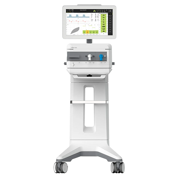
Modes and characteristics of the Elisa 800.
Special ventilation modes help reduce asynchronous triggering and improve weaning success rates. A comprehensive, modular, and optional system allows, for example, indirect bedside calorimetry, EEG-based sedation monitoring, bi-level nasal ventilation using cannulas, and automatic control of inspiratory O2 concentration.

Application of volatile anesthetics.
Daily awakening attempts, propofol infusion syndrome, rapid neurological assessment of ventilated patients in intensive care, and reduction of brief reactive psychosis—these are all reasons for using volatile anesthetics in intensive care. We decided to take on this challenge and implemented a comprehensive strategy for "essential safety and performance for anesthesia systems." This encompasses more than just the safe operation of intensive care ventilators and the effects of anesthetic gases on ventilator materials. The Anesthetic function compensates for inspiratory and expiratory resistances in the Anaesthetic Conserving Device (Sedaconda) system, thus preventing a prolongation of mean expiratory time, reducing the risk of air trapping, and ensuring accurate volume measurement.
In combination with the LeoLyzer multi-gas sensor, it is optionally possible to accurately measure and monitor anesthetic gases directly with an ELISA.

Cuffscout
Continuous monitoring and control of the blocked cuff are among the metry measures aimed at reducing the risk of ventilator-associated pneumonia (VAP) in ventilated patients in intensive care units. Intermittent cuff control, often used until now with a manometer, is insufficient to mitigate this risk. That's why we've equipped our successful products with the new "Cuffscout" function. This allows for maintaining and monitoring the cuff pressure prescribed by the user.
In addition, our devices immediately detect faulty cuffs and leaks, and feature an algorithm for cough detection. This further simplifies individual cuff adjustments.

Transpulmonary pressure monitoring
Lung-protective ventilation reduces ventilation-associated complications, notably by decreasing the mechanical pressure and volume load on the lungs. Knowledge accumulated in recent years has proven that lung-protective ventilation is only possible by regularly adjusting ventilator settings to individual lung function. But what happens if the standard guidelines for lung-protective ventilation can no longer be followed?
Adapting ventilation therapy based on transpulmonary pressure measurement is a simple, minimally invasive, and valid method that requires only the placement of a modified gastric tube. Changes in esophageal pressure during a respiratory cycle reflect changes in pleural pressure. As the difference between ventilatory pressure and pleural pressure, transpulmonary pressure indicates the extent of mechanical stress exerted on the alveoli, which is consequently responsible for ventilation-associated lung injury. The inspiratory plateau pressure set on the turbine plays a secondary role. Studies have shown that, due to the significant variability in the lung-thorax elasticity ratio, a set inspiratory plateau pressure on the ventilator results in very different transpulmonary pressure gradients. In patients with elevated pleural pressure, for example, due to high intra-abdominal pressure, the same inspiratory pressure can be achieved with less ventilator-associated lung injury than in patients with low pleural pressure. Expiratory transpulmonary pressure (eTPP exsp) can then be adjusted by titrating the applied PEEP, since airway pressure is related to the applied PEEP. Unlike other methods for detecting individual PEEP, this method can also be used during spontaneous breathing and weaning. During weaning, esophageal pressure measurement can provide valuable information (unmasking patient-ventilator asynchrony, monitoring respiratory muscle effort, calculating intrinsic PEEP during spontaneous respiration, etc.) and allows for optimization of the weaning process. In this context, it is possible to determine the patient's work of breathing during assisted spontaneous respiration in acute situations, so that the necessary support can be directly adapted to the patient's individual lung function through pressure-based assistance.

Volumetric capnometry.
In the era of lung-protective ventilation, ventilation efficiency can be optimized through targeted measurements of the dead space/tidal volume ratio. Capnography, as a graphical representation of expiratory CO2 concentration, is an essential component of bedside monitoring for ventilated patients. Capnography non-invasively and in real time represents CO2 kinetics. In daily routines, it primarily allows for the identification of correct intubation and adjustment of the minute respiratory volume. However, capnography, particularly in its volumetric form, which is not yet widely used in hospitals, can provide much broader and clinically valuable additional information. This includes monitoring and optimizing ventilation, as well as assessing gas exchange. The healthcare team thus obtains clinical parameters for bedside decision-making that, until now, could only be obtained through more complex, invasive, and non-automated procedures.

LEOCLAC.
Automatic regulation of inspiratory oxygen concentration based on pulse oximetry allows for oxygen delivery according to guidelines. High O2 concentrations can lead to unexpected events. The spectrum ranges from inflammatory airway reactions and resorption atelectasis to seizures and increased in-hospital mortality. During high-flow oxygen therapy and ventilation, oxygen saturation must be closely monitored, and the inspiratory oxygen concentration must be continuously adjusted to the individual therapeutic range. LEOCLAC, based on integrated pulse oximetry, allows for continuous adjustment of the inspiratory oxygen concentration to the defined therapeutic range. Compatible with invasive or non-invasive ventilation and high-flow therapy, LEOCLAC continuously evaluates pulse wave quality and detects potential artifacts. Various sizes and models of SpO2 sensors are available for LEOCLAC. Heart rate, oxygen saturation, and plethysmography waveform can be monitored independently by LEOCLAC. An intelligent graph facilitates the assessment of FiO2 regulation.

PEEPfinder.
It is well established that the ventilation-synchronous collapse and reopening of lung zones in patients with ARDS significantly damages lung tissue. In particular, ventilation-synchronous opening and closing (alveolar cycling) of lung zones represents an independent risk factor for increased mortality. The PEEPfinder can be used to optimize ventilator settings and thus assists in lung-protective ventilation. The maneuver is performed within a safe window and can be combined with a preoxygenation function. The extended quasi-static P/V Tool assists the user in stress assessment. Intelligent algorithms and comprehensive safety features allow for the easy determination of lung elastic properties. Extensive assessment options are available for this purpose. Graphical assessment support for detecting inflection points, collecting stress indices, and up to ten reference loop recording options simplify the implementation of ventilation.
Product features
High-flow oxygen therapy
Ventilation non invasive

Reviews
There are no reviews yet.